
Pathology & Cytology Services
Comprehensive histopathology with surgical tissue grossing, paraffin embedding, microtomy, special staining, and cytological evaluation. All specimens signed out by certified pathologists with formal multi-tier reviews.
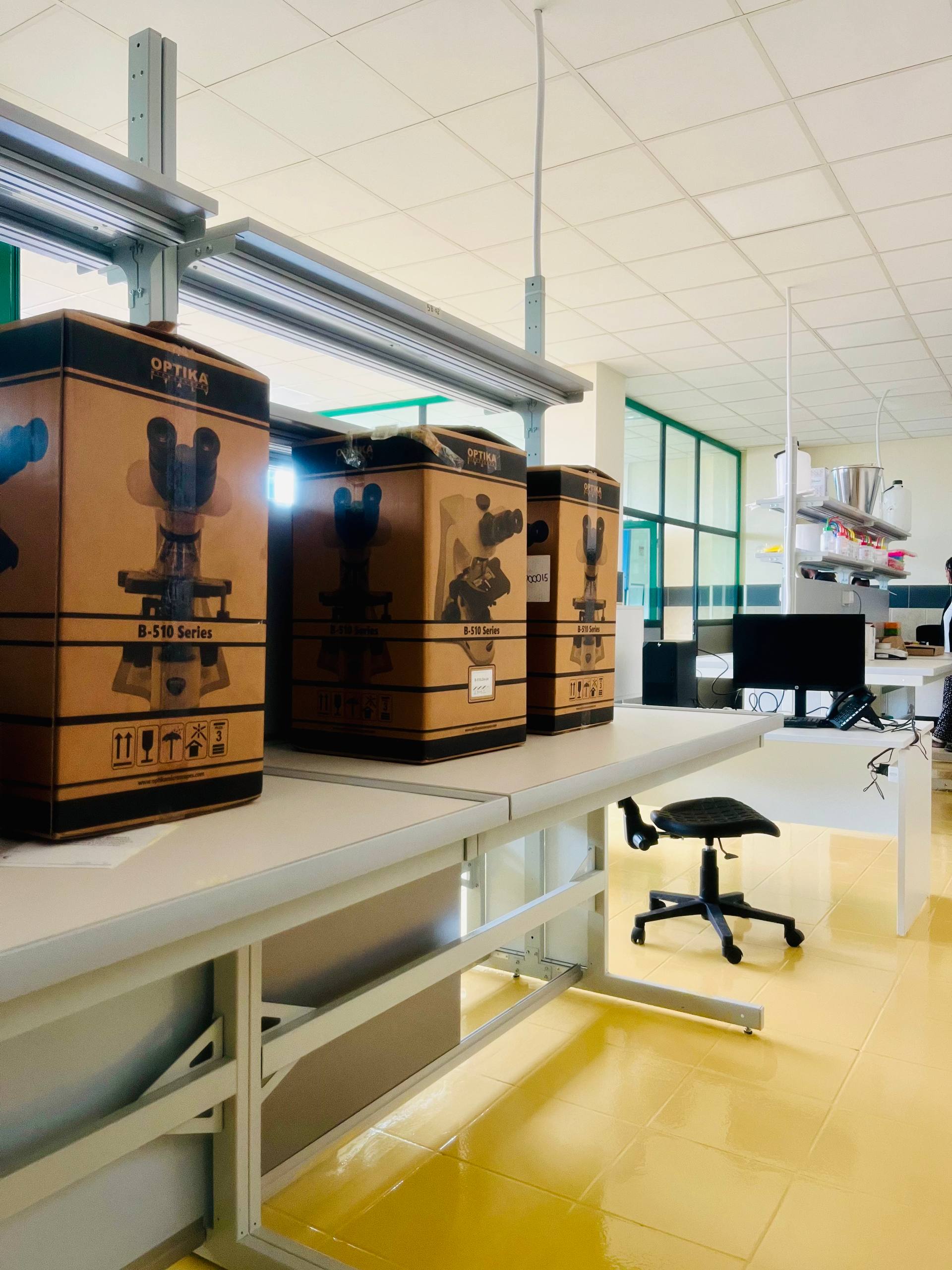
Advanced optical microscopy suite featuring binocular and trinocular research microscopes for cellular morphology and histology.
The Pathology and Cytology division provides comprehensive surgical tissue grossing, paraffin embedding, microtomy, special staining, and cytological evaluation. Every specimen is examined and signed out by certified pathologists with formal multi-tier diagnostic reviews for neoplastic and complex cases. The division integrates with the Molecular Division for reflex HPV genotyping on abnormal cervical cytology results, creating a seamless diagnostic pathway from cytology screening through confirmatory molecular testing.
Biopsy tissue in 10% neutral buffered formalin (container volume at least 10× tissue volume). FNAC air-dried and alcohol-fixed slides with accompanying clinical history.
Featured Instrumentation & Equipment Fleet
World-class clinical analyzers powering this division
Automated Tissue Processor & Microtomy Suite
Histopathology SuiteEnclosed vacuum tissue processing for consistent, standardized dehydration, clearing, and paraffin infiltration.
Rotary precision microtomy with automated temperature-regulated flotation baths.
- Uniform 3–4 micron ribbon sectioning for optimal nuclear and cytoplasmic morphology
- Standardized Hematoxylin & Eosin (H&E) staining protocols with strict pH control
- Temperature-controlled flotation water baths for wrinkle-free section mounting
Liquid-Based Cytology (LBC) Processor
Cytology DivisionBatch processing of liquid-based cervical samples for high-quality monolayer slide preparation.
Centrifugation and thin-layer deposition technology for superior cellular preservation.
- Superior cellular preservation compared to conventional smear techniques
- Residual LBC sample can be directly forwarded to Molecular Division for reflex HPV genotyping
- Reduced obscuring blood, mucus, and debris for clear morphological evaluation
Facility & Equipment Gallery
Authentic photographs of instruments, analytical bays, and laboratory infrastructure
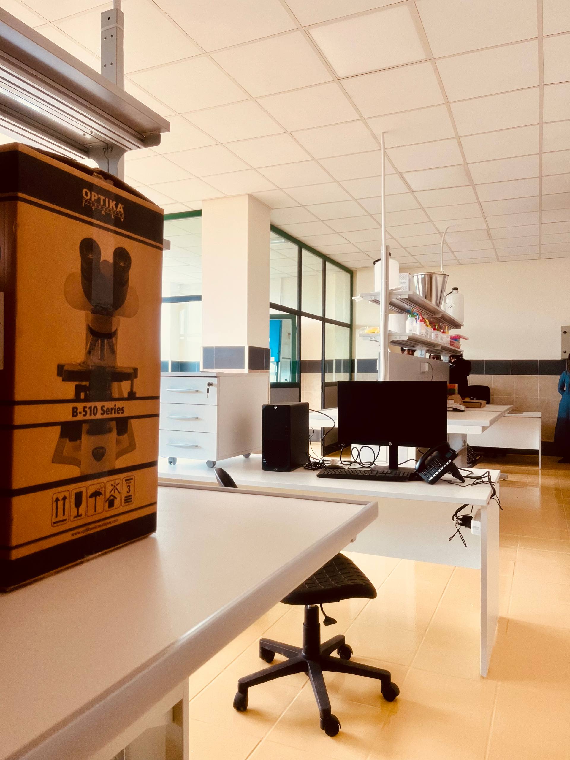
Morphology Review & Workstation Desk
Integrated diagnostic workstation for slide examination and pathologist sign-out review.
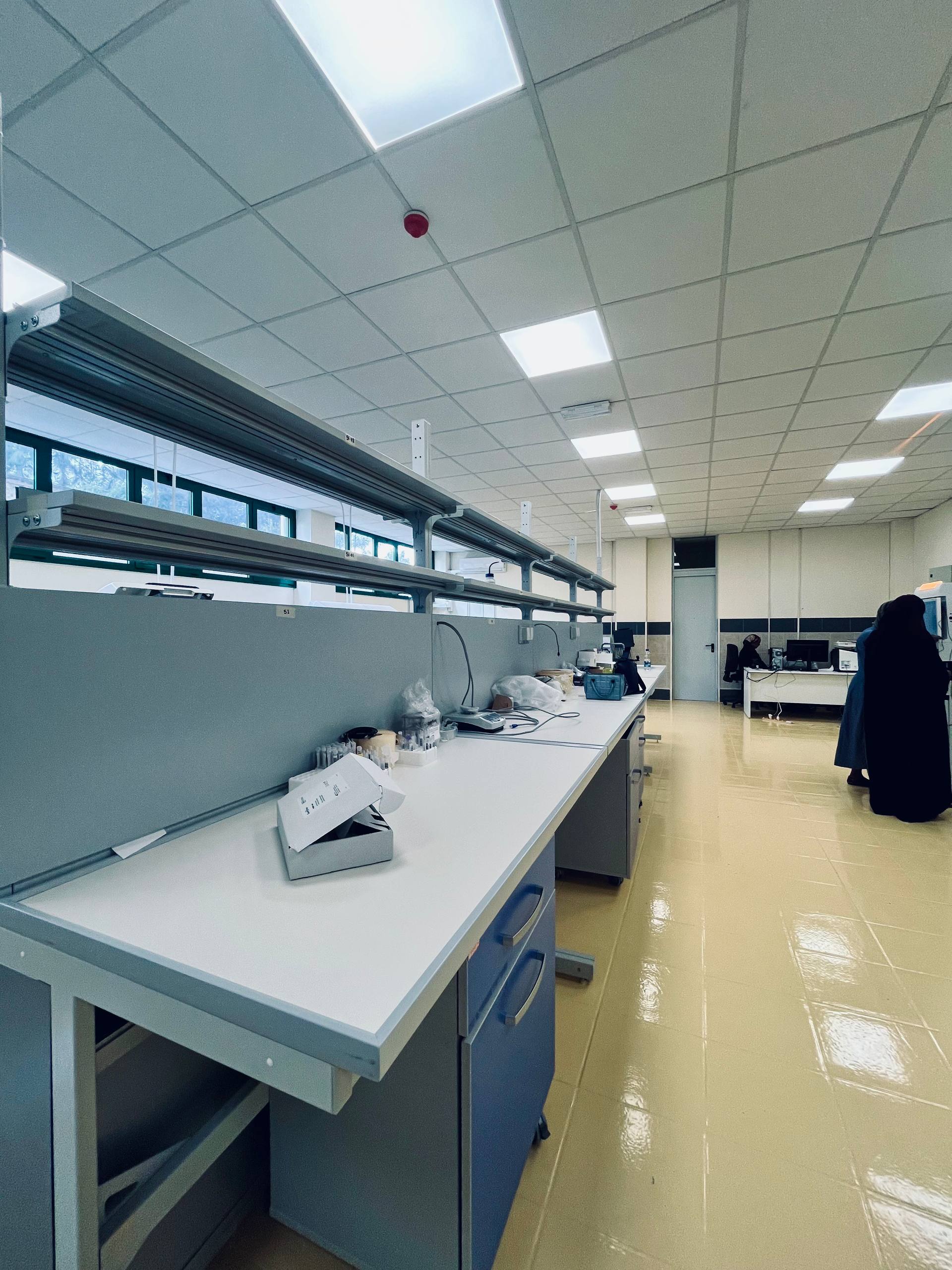
Analytical Staining & Preparation Benches
Spacious preparation rows for grossing, manual slide staining protocols, and special stains.
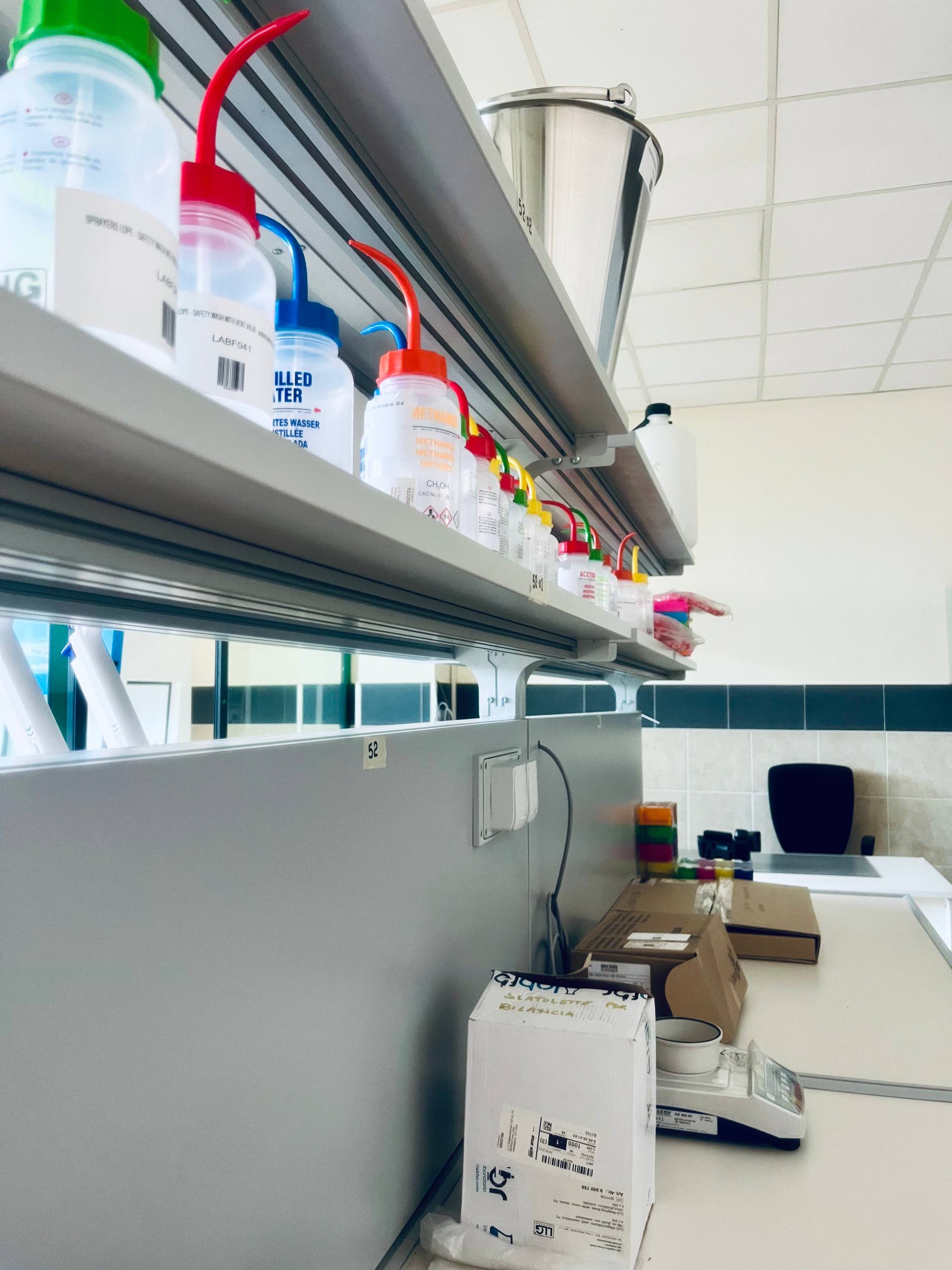
Analytical Weighing & Staining Solutions
High-precision analytical balance and color-coded safety wash solutions for specialized histology staining.
Clinical Assay Menu & Test Panels
Complete testing capabilities available in this division
Surgical Histopathology
- Surgical Biopsy — Gross Description, Microscopic Evaluation & Pathologist Report
- Endoscopic & Punch Biopsy Evaluations
- Resection Margins & Tumor Node Staging
- Special Histochemical Stains (PAS, GMS, ZN, Giemsa, Congo Red)
Exfoliative & Aspiration Cytology
- Fine Needle Aspiration Cytology (FNAC) with Rapid On-Site Evaluation (ROSE)
- Liquid-Based Cervical Cytology (Thin-layer Pap Smear)
- Pleural, Peritoneal, and Pericardial Fluid Cytology
- Urine Cytology for Urothelial Malignancy
- Bone Marrow Aspirate Cytology & Iron Stain Evaluation
Integrated Molecular Reflex Testing
- Reflex HPV Genotyping on Abnormal Cervical Cytology (LBC)
- Molecular Confirmation Following Suspicious FNAC
Analytical Advantages & Special Capabilities
Secondary independent pathologist review for all malignant diagnoses
Integration with Molecular Division for reflex HPV genotyping on abnormal cervical cytology
Rapid On-Site Evaluation (ROSE) for FNAC adequacy assessment during procedure
Dual-pathologist sign-off protocol for all oncological case reporting
MLT/MLS Student Hands-On Training
MLT/MLS students at ECUSTA rotate through the Pathology and Cytology laboratory, learning tissue grossing, sectioning, H&E staining, and cytology slide preparation under the mentorship of certified pathologists. Students also participate in quality assurance review processes and understand the integrated workflow between cytology findings and molecular diagnostic confirmation.

Request Testing from This Division
Submit an inquiry for Pathology & Cytology testing, or speak with our clinical coordinator for custom panel design and assay feasibility evaluation.